Tardimap is a global scientific collaboration building the first complete wiring diagram of a tardigrade's nervous system, reconstructing every neuron and connection in a whole animal.
Tardigrades have a compact nervous system of only a few hundred neurons, yet they coordinate complex eight-legged locomotion and behaviour. As panarthropods, they offer a rare window into the origins of neural processing, and a nervous system small enough to reconstruct in its entirety. A complete tardigrade connectome will be a foundational resource for invertebrate neurobiology and the neural basis of behaviour.
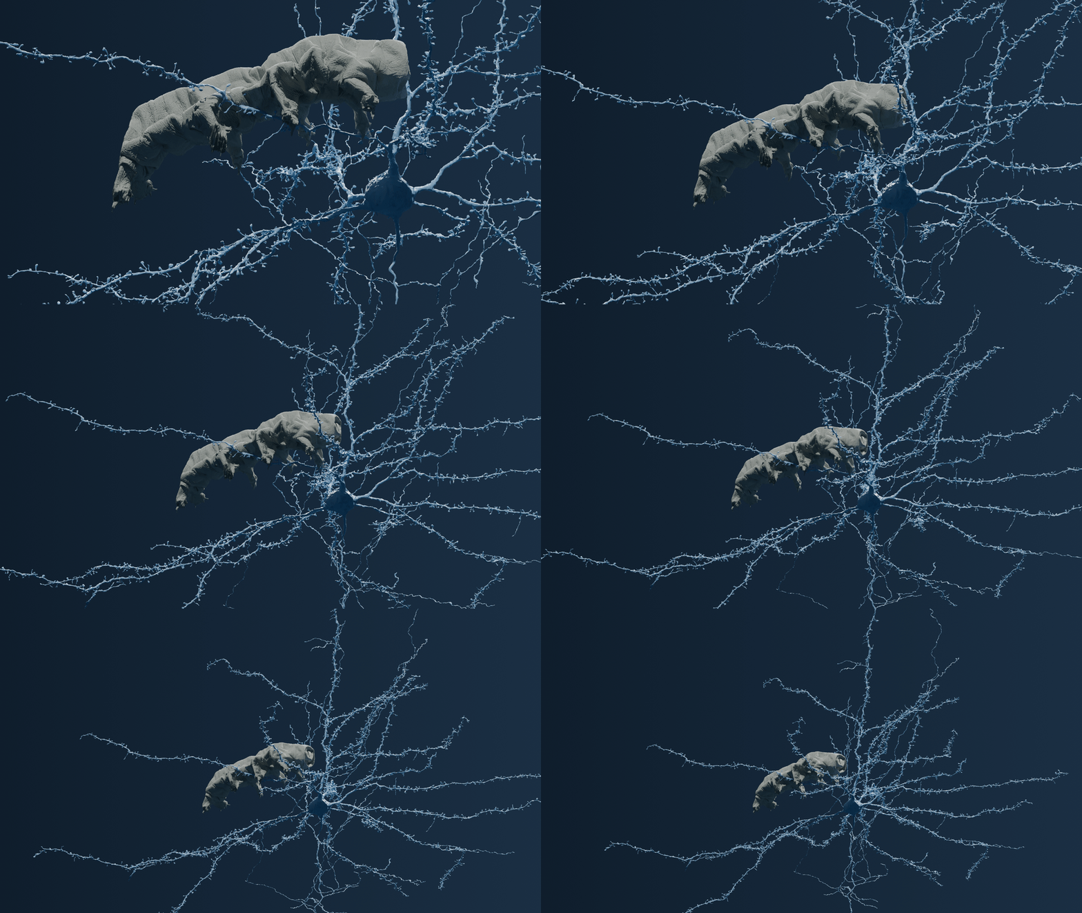
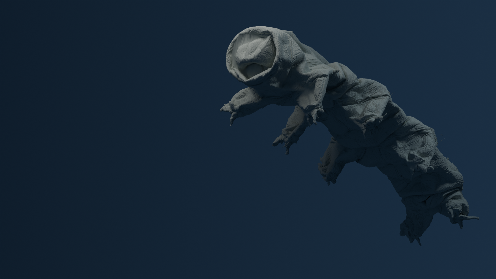
A whole tardigrade, shown to scale beside a single mouse pyramidal neuron (rendered from the MICrONS dataset).
Inside the animal
See the data come to life
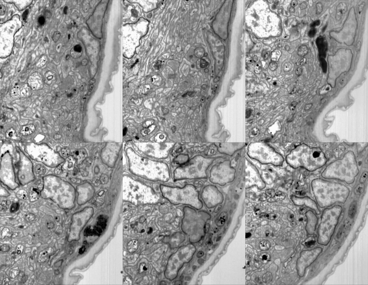
Electron microscopy lets us see nervous tissue at nanometer resolution, down to individual synapses.
Inside the brain: FIB-SEM of the neuropil.
Flythrough of the whole Riker EM volume.
Our method
How do you map the nervous system of a tardigrade?
From living animal to open dataset, in four stages.
01
Image
We image a whole tardigrade in ultra-thin slices with electron microscopes, down to the nanometer.
02
Reconstruct
We trace every neuron and connection through the slices to rebuild the nervous system in 3D.
03
Model
We turn the wiring diagram into models of how circuits drive behaviour.
04
Share
All data, tools, and results will be released openly for the research community.
Meet Riker, our first whole-body dataset
The first complete whole-body electron-microscopy dataset of an adult Hypsibius exemplaris. Imaging is complete; reconstruction is underway.
Tardigrades are microscopic, eight-legged invertebrates that live in diverse environments ranging from deep oceans to damp moss. They are widely known for their ability to enter a dormant state called cryptobiosis, allowing them to survive extreme temperatures, high pressure, radiation, and the vacuum of space. Once reintroduced to water, they rehydrate and return to their normal active state.
H. exemplaris walking under a polarised-light microscope.
Tardigrades possess a segmented body plan consisting of a head and four trunk segments, each supporting a pair of legs. Despite having a relatively small number of cells and a simple nervous system with few neurons, they exhibit complex limb coordination and can adjust their walking gaits based on the surface they are traversing. They also feature rudimentary eyespots, known as ocelli, which allow them to detect light.
Over 1,300 species of tardigrade have been described, inhabiting nearly every environment on Earth — from Antarctic ice sheets to tropical rainforests and deep-sea sediments. While many share the hallmark traits of cryptobiosis and radiation resistance, their exact physiological and neurological properties vary considerably across the phylum. Our research focuses on Hypsibius exemplaris, a freshwater species that has become the leading laboratory model for tardigrade biology thanks to its transparency, short generation time, and well-characterised genome.
The roadmap
Towards a whole-body connectome
We present the first whole-body electron-microscopy dataset of an adult Hypsibius exemplaris. Our goal is to reconstruct complete whole-body connectomes, mapping every neuronal connection and how neurons interact with non-neuronal tissue, to understand how a simple nervous system coordinates complex behaviour.
Phase 0
Initial challenges
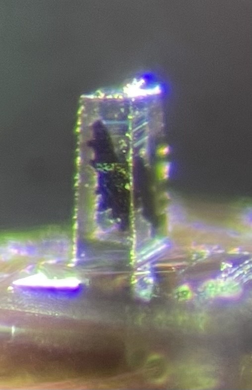
The tardigrade's resilience and its ability to rapidly enter cryptobiosis make sample preparation difficult. The volumes presented here were stained with heavy metals to preserve ultrastructural integrity for high-resolution electron microscopy.
A plastic-embedded tardigrade, prepared for EM.
Phase 1
High-resolution imaging
Using multibeam scanning electron microscopy (multiSEM) and focused ion beam scanning electron microscopy (FIB-SEM) to capture complete volumetric data of the nervous system at synaptic resolution.
FIB-SEM volume of the tardigrade brain.
Phase 2
Neural reconstruction
Employing image-segmentation algorithms and machine learning to trace individual neurons, processes, and synaptic connections throughout the entire nervous system.
Phase 3
Comparative analysis
Comparing connectomes across multiple tardigrade datasets to investigate variability between individual animals.
We are building open, high-resolution whole-body datasets of Hypsibius exemplaris. Each entry below is a distinct imaging acquisition.
Riker
Hypsibius exemplaris
FIB-SEMImaging completeReconstruction in progress
The first complete whole-body electron-microscopy dataset of an adult tardigrade, imaged at 8-nanometer isotropic resolution.
Sample prep done by Nelson Medina, eFIB-SEM imaging done by Nelson Medina and Marc Corrales, segmentation done by Nelson Medina and Anastasia Sorokina.
What we have
A fully imaged and aligned 3D volume containing the complete nervous system, musculature, and organ systems of Hypsibius exemplaris.
Flythrough of the whole electron-microscopy volume.
Ongoing — segmentation
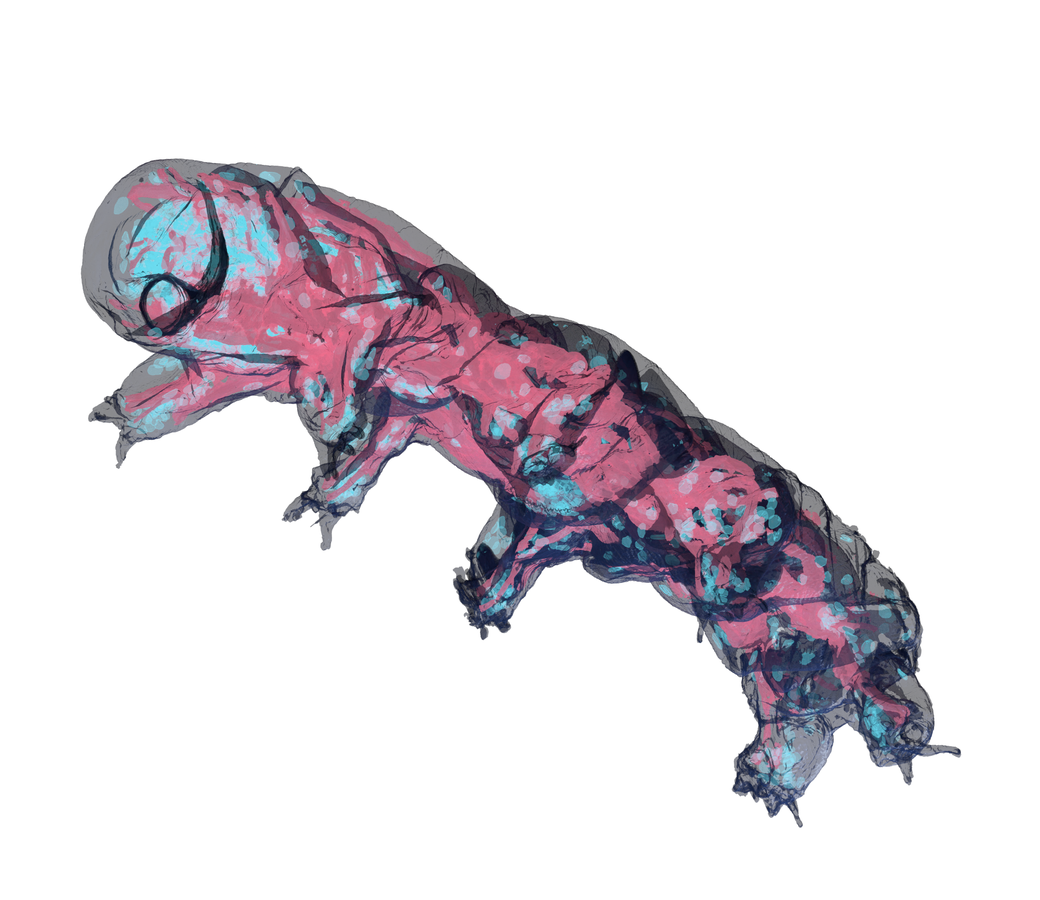
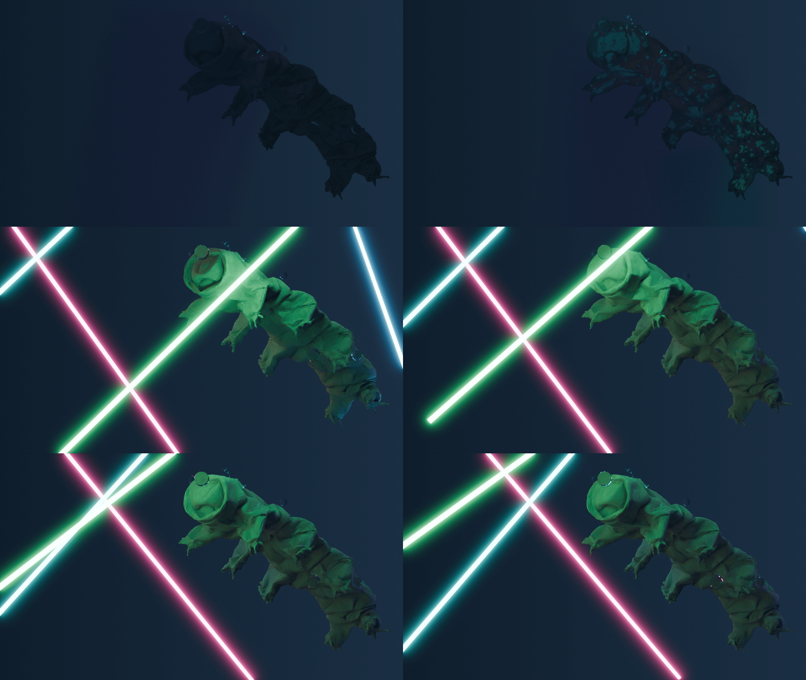
Initial segmentation and neural tracing are underway, with early reconstructions revealing the architecture of the brain and ventral nerve cord. Select a layer to highlight it in the 3D reconstruction.
SynapsesMitochondria
Segmentation layers of the whole-body EM dataset (Riker).
What's next
Complete neural reconstruction and synapse identification.
Quark
Hypsibius exemplaris
multiSEMImaging completeComing soon
Details coming soon.
Dataset C
Hypsibius exemplaris
TBDComing soon
Details coming soon.
About Tardimap
Tardimap began in the Kornfeld lab at the MRC Laboratory of Molecular Biology, where the first whole-body tardigrade volumes were imaged. The project is led by Anastasia Sorokina and Joergen Kornfeld, and has grown into a global collaborative effort bringing together different areas of imaging and biology expertise.
Our team (A–Z)
A global network of imaging scientists, connectomics researchers, and neuroscientists.
Interested in the project? We're always glad to hear from researchers and potential collaborators. Use the form below to introduce yourself and tell us a little about your interests, and we'll be in touch.